DGP Annual Conference 2023: Unleashing the Power of Digital Diagnostics and Virtual Reality
Written by Somaia Abubaker on 05.07.2023
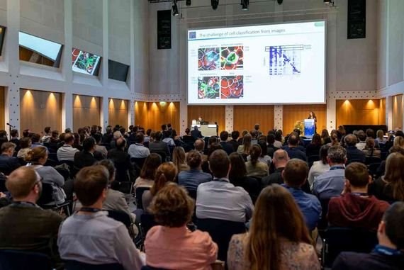
As part of our ongoing project med4PAN, our team member Ilgar Guseinov attended the Deutsche Gesellschaft für Pathologie (106th) Annual Conference, which is organized by Prof. Falko Fend and his staff from the Institute of Pathology at the University Hospital in Tübingen. The conference took place in Leipzig, Germany. The main topics discussed were related to hematopathology, multiparametric cell and tissue analysis, intraoperative tissue sensors, pathology and interdisciplinarity. The conference consisted of joint meetings with other professional societies as well as meetings on all areas of pathology.
Our ongoing project as part of the 5G-Innovationsprogramm funded by Bundesministerium für Verkehr und digitale Infrastruktur (BMVI) has one use case on digital histopathology. We aim to develop a remote diagnostic methodology for pathologists, by which pathologists in a specific location can use a web camera configured to a microscope. This setting is connected to computer’s software designed to display the specimen on screen and share it with another pathologist from a different location through a video conferencing platform. Another method we are working on is using the Whole Slide Scanner (WSI) to take more comprehensive shots of the specimen, enhancing the accuracy and reliability. Thus, pathologists can help in the primary and secondary diagnosis for specimens remotely either in a synchronous or asynchronous manner.
A lot of abstracts discussed during the conference were interesting in relation to our project. Dr.Kinzler, at University Hospital Frankfurt discussed the Fluorescence Confocal Microscopy (FCM) on Liver Specimens for Full Digitization of Transplant Pathology Using Vivascope 2500-G4, which is a confocal laser scanning microscope designed for the analysis of diagnostic biopsies, and the assessment of tumor margins. He compared the process of assessing tumor margins in liver biopsies between the FCM and the conventional histology. Findings suggest almost perfect agreement as well as shorter scanning time. Limitations are due to the delayed legislation of the device. Prof. Litjens, at the Radboud University Medical Center, discussed the project The open source whole slide imaging IO library and viewer. IMI BIG PICTURE has a vision to create the first European GDPR compliant platform, in which quality controlled WSI data and advanced AI algorithms will exist. Other projects in relation to artificial intelligence and pathology in prostate cancer identification were also discussed.
DGP Annual Conference 2023Prof.Dr.Schüffler, from Technical University Munich (TUM), presented the existing lab in TUM for cancer detection, segmentation and quantification in digital histological slides. He mentioned important points in terms of the appropriate integration of AI into the pathologist’s workflow, regulatory considerations, and the importance of validating AI tools by adequate studies. We are in contact with the professor for further discussion of our ongoing project. Prof.Jukouka, at Nagasaki University-Japan, discussed the digital transformation of the pathology laboratory at Kameda medical center in Japan. They created a digital pathology network to exchange expertise regarding consultation and education. We are inspired by their transformative journey, and we are interested in understanding the guidelines they adapted to establish the network effectively. Talking to top-notch experts in pathology, oncology and computational sciences gives us a comprehensive approach to the development of digital pathology.
We were pleased to introduce our med4PAN project progression and ideas to other fellows interested in the field of pathology, and digital histopathology. We discussed various aspects in terms of the difficulties of the regulation of such practices, and suggestions to overcome them so that such technologies can be effectively used in practice in the near future. Professionals discussed challenges they encounter while working on their studies. Challenges are not limited to usability and reliability tests, but also to ethical and legal regulation, marketing, and sustainability. Experts in the domain confirmed that it is not sufficient to have a well-equipped laboratory, it is crucial to develop working protocols and operating procedures. This is especially important for experts working remotely in home offices. The ongoing studies give a promising future for digital pathology.
The conference gave us the boost necessary to go on with our project. It also embedded some aspects worth focusing on while developing our model. As we are working on the use of Whole Slide Scanner devices for the proper primary diagnosis of various specimens in histopathology domain, specifically oncology, we are further interested in incorporating Virtual Reality (VR) glasses into the viewing of the specimens by pathologists to yield a better immersive experience as well as better visualization and interpretation of the slides, diagnosis, sub-diagnosis, and recommendations.

